
The clinical management of an intervertebral disc herniation—commonly referred to by patients as a slipped disc, bulging disc, or ruptured disc—has undergone a radical paradigm shift over the last decade. Modern medicine has transitioned from reactive surgical interventions toward a comprehensive, multi-modal rehabilitation framework.
As a leading cause of global musculoskeletal disability affecting millions annually, disc-related pathologies require a nuanced understanding of spinal anatomy, biomechanics, and the lifestyle factors that trigger neural irritation.
1. The Biomechanical Foundation: Anatomy and Risk Factors
The human spine is a marvel of biological engineering. Central to its function is the intervertebral disc, which acts as the primary shock absorber between your vertebrae. To understand a herniation, it helps to picture a “jelly doughnut”:
- Nucleus Pulposus (The Jelly): The gelatinous inner core (80% water) that distributes axial loads.
- Annulus Fibrosus (The Dough): Concentric collagenous rings designed to contain the nucleus and resist tensile stress.
As the disc ages, it undergoes desiccation (loss of water content), leading to micro-tears in the annulus. A herniated disc occurs when the inner “jelly” pushes through a crack in the tough outer layer.
Key Risk Factors & Causes:
- Demographics: Most common in men aged 30 to 50.
- Lifestyle: Smoking (which reduces oxygen supply to the disc), obesity, and sedentary habits.
- Biomechanics: Improper lifting techniques, repetitive twisting, and poor posture.
2. Pathophysiology: Mechanical Compression vs. Biochemical Irritation
A herniated disc is rarely just a structural failure. Symptoms are governed by two interrelated mechanisms:
- The Biochemical Shift: Modern research emphasizes that pain is often triggered by a “chemical storm.” When the inner disc material is exposed to the immune system, it releases pro-inflammatory cytokines. These sensitize nerve roots, causing severe pain even without significant physical compression.
- Stages of Disc Displacement:
- Bulge: The annulus remains intact but weakens, causing localized stiffness.
- Protrusion: The nucleus pushes into the inner rings.
- Extrusion: The nucleus breaches the outer annulus, often leading to sharp radiculopathy.
- Sequestration: A nuclear fragment separates from the disc entirely.
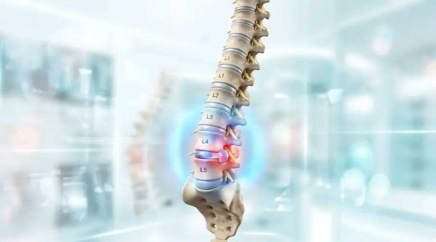
3. Segmental Analysis and Clinical Manifestations
The location of the herniation dictates the specific symptoms. Although they can occur anywhere, they are extremely rare in the mid-back (thoracic spine):
- Lumbar Spine (L4-S1): The most frequent site of injury. Symptoms include lower back pain, muscle weakness, and Sciatica—a sharp, burning pain radiating from the buttocks down the leg to the foot.
- Cervical Spine (C5-C7): Results in neck pain (especially when turning the head), weakness in the arms, and tingling/numbness radiating into the shoulder blades, hands, or fingers.
4. The Diagnostic Algorithm: Beyond the MRI Paradox
A common clinical pitfall is over-reliance on MRI. Studies show that a large percentage of asymptomatic individuals have disc abnormalities on imaging. Elite diagnostic protocols now include a combination of tools:
- Clinical Correlation: Matching physical symptoms with provocative tests like the Straight Leg Raise (SLR).
- X-Rays & CT Scans: While plain X-rays cannot detect a herniated disc, they are crucial for ruling out tumors, infections, or broken bones.
- Electrodiagnostic Testing (EMG/NCS): Measures electrical impulses to pinpoint exactly which nerve root is damaged and assess the severity of neural compromise.
- MRI: Used to confirm the exact location and size of the herniation when conservative treatments fail.
5. First-Line & Integrated Conservative Management
Approximately 90% of herniated discs can be successfully managed without surgery within a few weeks to months.
A. At-Home Care & Medication
- Activity Modification: Avoid heavy lifting and prolonged sitting. Crucially, avoid prolonged bed rest (more than 1-2 days), as it causes muscle stiffness. Gentle movement is key.
- Thermal Therapy: Applying cold packs initially to reduce swelling, followed by heat therapy to relax muscle spasms.
- Pharmacology: Over-the-counter NSAIDs (Ibuprofen, Naproxen) for inflammation. In severe cases, doctors may prescribe muscle relaxants or neuropathic drugs (like Gabapentin) for nerve pain.
B. Advanced Rehabilitation & Technology
- The McKenzie Method: Focusing on “Centralization”—moving the pain from the extremities back toward the spine.
- Non-Surgical Spinal Decompression: A computer-controlled technology creating negative intradiscal pressure. This “vacuum effect” encourages the retraction of herniated material and nutrient diffusion.
- Reformer Pilates: Unlike traditional exercises, Reformer Pilates uses a spring-based system to provide eccentric control. This strengthens the “natural corset” (transverse abdominis) without imposing shear forces on the vulnerable disc.
C. Epidural Steroid Injections (ESIs)
If oral medications and therapy plateau, a targeted injection of corticosteroids directly into the spinal space can drastically reduce nerve inflammation and provide a window of pain relief for physical therapy.
6. When Surgery is Mandatory: Surgical Options & “Red Flags”
While conservative care is the gold standard, surgical intervention becomes necessary if a patient experiences “Red Flag” symptoms:
- Cauda Equina Syndrome: Emergency loss of bladder/bowel control and “saddle anesthesia.”
- Progressive Motor Weakness: Such as “Foot Drop.”
- Intractable Pain: Unresponsive to all conservative measures after 6 weeks.
Modern Surgical Techniques:
If surgery is required, minimally invasive options are preferred:
- Microdiscectomy: Using a microscope to remove only the protruding portion of the disc that is pressing on the nerve.
- Laminectomy: Removing a small portion of the vertebral bone (lamina) to open up the spinal canal and relieve pressure.
7. Prevention and Long-Term Spinal Hygiene
Preventing recurrence is a lifelong commitment. Key preventative measures include:
- Proper Lifting Mechanics: Bend at the knees, not the waist. Keep loads close to the body.
- Ergonomics & Posture: Avoid slouching, take frequent stretching breaks if you have a desk job, and avoid wearing high heels.
- Weight Management & Diet: Reducing excess weight to lower the mechanical load on the lumbar spine.
Final Conclusion
Modern disc management is a story of patient empowerment. By integrating precise diagnostics, lifestyle modifications, non-surgical technologies like spinal decompression, and biomechanically sound movement such as Pilates, patients can achieve a sustainable recovery. The goal is no longer just symptom relief, but restoring the body’s natural resilience.
If you are experiencing back pain or suspect a herniated disc, timely intervention is critical. Explore our expert Physiotherapy and Chiropractic services, or book a consultation today to begin your personalized recovery protocol.